In recent years, optical coherence tomography (OCT) has become the standard of care for retinal disease diagnosis and is now widely used for coronary artery disease and intravascular imaging. Sustained innovations have driven commercial modalities – such as swept-source (SS-OCT) – to speeds of 100,000-400,000 sweeps per second. That pace shows no sign of slowing, with OCT’s trajectory reflected in its inclusion under the Biomedical Applications sub-committee at CLEO 2026 – a clear signal of where the photonics community’s attention is focused.
The shift from time-domain (TD-OCT) to Fourier-domain OCTs – including SS-OCT and spectral-domain OCT (SD-OCT) – was a technological turning point for diagnostic imaging. In unlocking Fourier-domain OCT, simultaneous depth profiling, enhanced sensitivity, and faster acquisition resulted in the ability to generate three-dimensional volumetric images for the first time – and development hasn’t plateaued. SS-OCT has reached megahertz-range (MHz) A-scan rates in research and surgical prototype contexts, with resolution and imaging depth advancing alongside speed.
From Retina to the Operating Room
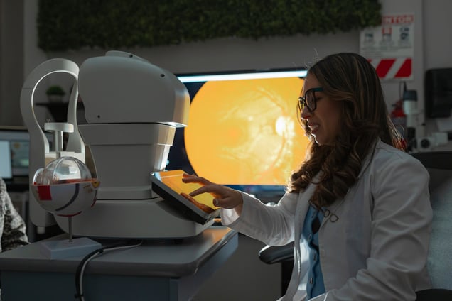
Although ophthalmology remains the dominant clinical application – diagnosing and monitoring retinal disease, age-related macular degeneration, and glaucoma – OCT is expanding across other medical specialties.
In cardiology, intravascular OCT enables vessel wall assessment, stent optimization, and plaque characterization – a capability progressing toward neurovascular territory. Meanwhile, dermatology and intraoperative surgical guidance are active areas of development, alongside endoscopic OCT.
In each clinical setting, the performance threshold is raised, demanding higher speeds for surgical guidance, finer resolution within compact catheter designs for cardiology, and a wider field of view (FoV) for dermatology.
Why Optical Component Precision is Imperative
Evolving performance gains and newfound applications for OCT depend on high-precision optical components that meet increasingly stringent spectral specifications. In SD-OCT, for example, optical properties determine whether the system achieves its theoretical resolution limit, with spectral uniformity, channel isolation, and wavelength-mapping accuracy as component-level constraints that carry direct diagnostic performance consequences.
In practice, spectral non-uniformity in SD-OCT spectrometry degrades resolution at the exact depths of deeper tissue features – often where pathologies reside – and as the technology extends into fields such as intraoperative applications, neurovascular imaging, and oncology, tolerance for any form of error at the optical component level further narrows.
The precision requirements this places on optical components – and how diffraction gratings and discrete patterning address them – are explored in more detail in our companion piece here.
OCT’s next development phase is being shaped by two advances: AI and miniaturization. AI-assisted image analysis is enhancing diagnostic accuracy and automating the identification of early-stage pathological markers, while increasingly compact systems are improving accessibility, extending OCT’s reach to point-of-care and remote deployment settings.
As OCT continues to advance into new clinical domains, the precision of every optical component becomes increasingly consequential. At Torrent Photonics, we fabricate stripe filters, micropatterned arrays, and transmissive gratings and precision optics that help OCT system integrators bridge the gap between specified performance and operational reality.
To discuss how we can support your work in OCT, contact our team today.