Medical imaging instruments increasingly rely on optical techniques, but their diagnostic value rests on the high precision of the optical components inside them.
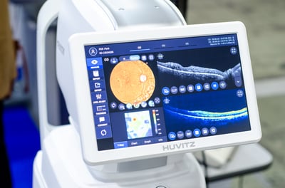
Alongside X-ray and MRI, optical imaging equipment – which uses light rather than radiation – is used to diagnose disease, monitor treatment, and guide procedures. Endoscopy, diagnostic modalities such as optical coherence tomography (OCT), and infrared (IR) imaging such as thermal imaging for tissue assessment all rely on light-based optics to provide clinical detail safely and in real time. That performance, however, is decided before light ever reaches the sensor.
How precision optics shape clinical imaging
Each optical technique places its own demands on components inside a system. In endoscopic procedures, an endoscope images the body from inside. It does this via a tube that’s only a few millimeters wide, meaning tiny objective gradient-index (GRIN) lenses, rod-lens relays, and prisms must all be held to sub-micron centration and surface figure within a fraction of a wavelength. Any error a larger lens would absorb becomes significant at this smaller scale. That is because diffraction-limited spot size does not shrink along with the lens: a few microns of decenter that would be lost in the blur circle of a 10 mm lens becomes a meaningful fraction of the whole aperture in a 2 mm one.
Meanwhile, clinical tools such as OCT, slit lamps, and surgical microscopes depend on optics that deliver resolution and light throughput to ensure fine structures, such as small vessels and early lesions, are discernible. Here, image quality feeds directly into what a clinician can detect.
In OCT specifically, axial resolution is set by the source bandwidth, so the optical train has to preserve that bandwidth without introducing a dispersion mismatch between the sample and reference arms. Thermal imaging for wound assessment, perfusion, and inflammation monitoring requires IR-transmitting materials. These technologies predominantly work in the long-wave IR (LWIR, roughly 8-14 μm), where ordinary glass is opaque, so specific substrates, such as chalcogenides, are needed.
Why aberration correction defines image clarity
Optical aberrations are systematic imperfections that distort how a lens focuses, resulting in degraded image clarity and color accuracy.
The most relevant to medical imaging are:
- Spherical aberration: occurs when rays striking the lens edge focus at a different point than rays passing through the center. This softens fine detail across the image, reducing contrast and sharpness.
- Chromatic aberration: occurs when different wavelengths focus at separate points. This shows up as color fringing at high-contrast boundaries, such as vessel edges, degrading the accuracy of fine detail.
Two component types address these aberrations:
- Aspheric lenses: use a non-spherical surface form to compensate for spherical aberration within a single lens.
- Achromatic lenses: combine two glass types of differing dispersion to bring multiple wavelengths to a common focus, correcting chromatic aberration.
Spherical and chromatic aberration is not the only factor worth tracking here. Coma, astigmatism, and field curvature tend to dominate in wide-field endoscopic and surgical optics, since these systems image well off the optical axis. Correcting them usually takes more than a single asphere or achromat, which is one reason distal-tip and wide-field designs often carry more elements than the two aberrations above would suggest on their own.
While optical specification matters, fabrication quality plays an equally decisive role. Surface-figure tolerance (in most cases, λ/10 or better) and the uniformity of coatings – the latter typically applied for anti-reflection or wavelength-selective functions – must be maintained to preserve the correction the design specifies. That figure is usually given as peak-to-valley at 633 nm; distal-tip endoscopic optics often need tighter control, sometimes specified as RMS rather than peak-to-valley, since a single high spot anywhere on the surface degrades the image.
How aspheric lenses enable compact systems
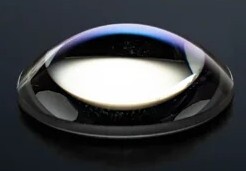
Aspheric lenses correct spherical aberration in one element instead of several, using the same correction principle covered above. For medical imaging devices specifically, this results in fewer elements that reduce size, weight, and power (SWaP) and is critical for handheld instruments, point-of-care systems and distal-tip optics. Fewer air-glass interfaces also reduce stray light and back-reflections.
Precision optics for medical imaging OEMs
Across these applications, optical precision determines whether equipment delivers its as-designed performance and meeting that standard comes down to fabrication.
At Torrent Photonics, we manufacture custom optical components – including aspheres, achromats, and prisms – and coated optics in visible and IR materials. That includes managing tolerance stack-up across a multi-element build and the metrology needed to confirm finished parts meet the design intent, not just the individual component prints.
To discuss the optical requirements for your application, please contact our technical team.
Image credit: Lutsenko_Oleksandr / Shutterstock.com